Abstract
A systematic review of 14 studies evaluated PRP as an adjunct in canine orthopedic procedures, finding safety and biological activity but limited evidence for consistent long‑term benefits. A concurrent experimental study on cranial cruciate ligament repair in 24 dogs demonstrated that PRP significantly improved complication scores and histological outcomes. Together, these studies suggest PRP is a promising biological adjuvant, while emphasizing the critical need for standardized preparation protocols.
Part I: The Systematic Review—What 14 Studies Tell Us
Vidal‑Negreira, García‑González, Valiño‑Cultelli, and González‑Cantalapiedra published a systematic review in Gels. Following PRISMA guidelines, the authors comprehensively searched PubMed, Scopus, and Web of Science, ultimately including 14 eligible studies—six experimental in vivo and eight clinical investigations—covering fracture repair, osteotomy, and tendon or ligament reconstruction.
What the review established:
– Safety: PRP demonstrated safety across all 14 studies, with no serious adverse events reported
– Early biological activity: Consistent early‑phase improvements in tissue regeneration and inflammatory modulation
– Gel formulation advantages: Gel PRP showed practical advantages in handling, local retention, and sustained growth factor release, acting as a transient fibrin scaffold
What the review left unresolved:
– Long‑term functional outcomes (months or more) were often similar to controls
– The marked heterogeneity in PRP preparations and outcomes across studies limits the strength of conclusions
– Optimal platelet concentration, injection frequency, and administration route remain undetermined
Methodological quality—assessed using ARRIVE 2.0, CONSORT, and RoB 2.0 guidelines—was moderate, with frequent omissions in blinding, sample‑size calculation, and preregistration. More than half of the studies failed to report fundamental parameters such as platelet concentration and leukocyte content.
Part II: The Experimental Study—PRP in CrCL Repair
Daradka et al. published a preliminary experimental study in Veterinary World. Cranial cruciate ligament injuries are a prevalent orthopedic issue in dogs, typically managed through surgical interventions such as tibial plateau leveling osteotomy. However, these techniques have limitations, including high costs and extended recovery periods.
Study design: Twenty‑four healthy male dogs were divided into two groups:
– Group A: surgery without PRP
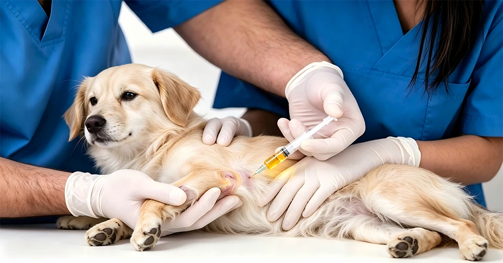
– Group B: surgery with intra‑articular PRP
The technique employed an autologous lateral digital extensor muscle tendon graft, and outcomes were evaluated through clinical assessments of lameness, post‑operative complications, and histological analysis at 10, 20, 30, and 40 days.
Results:
– Post‑operative complication scores: PRP group significantly better (p = 0.0025)
– Histological outcomes: PRP group significantly better (p = 0.0002)
– Graft maturation: unaffected by PRP but improved over time (p = 0.0013)
– PRP‑treated dogs exhibited faster recovery, enhanced tissue regeneration, reduced inflammation, and improved graft‑bone attachment
Interpretation: PRP improved the host’s response to surgery—the early healing environment, tissue regeneration, and graft‑bone integration—but did not directly alter the graft’s intrinsic maturation process. In this context, PRP functions as an “environmental modulator” rather than a “repair agent” for the graft itself.
Connecting the Two Evidence Streams
The systematic review found that long‑term functional outcomes often did not differ from controls. The experimental study found that PRP improved early healing but did not affect graft maturation. Both findings point in the same direction: PRP is a useful adjunct for early‑phase healing, but its role is modulatory rather than curative. It improves the conditions under which healing occurs, rather than directly driving the repair of damaged structures.
This distinction has practical implications for clinical expectations. PRP in orthopedic surgery should be positioned as a tool to optimize the healing environment—reducing inflammation, enhancing tissue regeneration, improving graft‑bone attachment—not as a guarantee of superior long‑term functional outcomes.
The Standardization Imperative
Both the systematic review and the experimental study highlight the same critical need: standardized PRP preparation. Without consistent preparation protocols—controlling for platelet concentration, leukocyte content, and activation status—the field cannot generate the reproducible evidence needed to establish clear clinical guidelines. The heterogeneity that currently limits the strength of conclusions is, at its root, a problem of preparation variability.
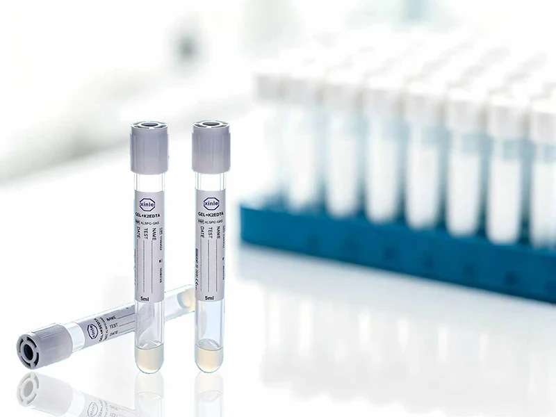
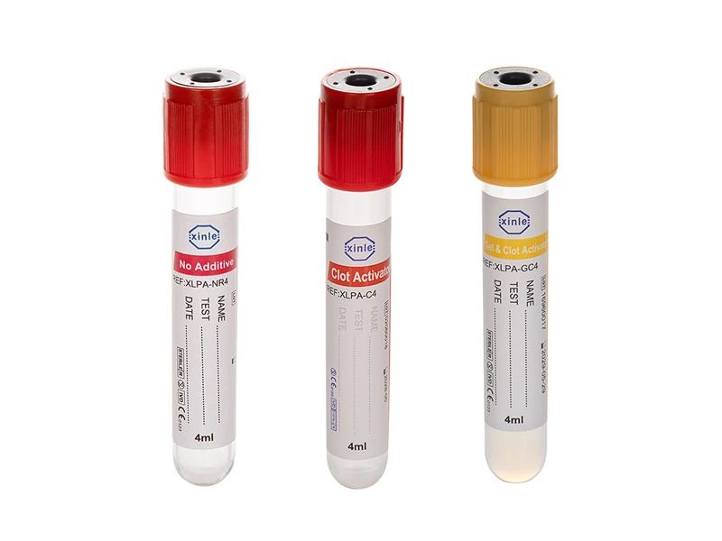
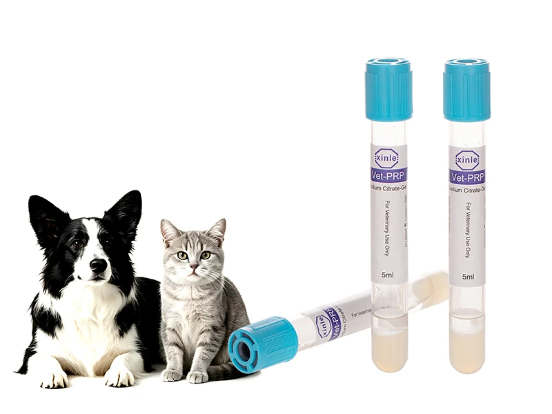
A standardized PRP preparation system—one that delivers consistent platelet recovery and purity across batches—addresses this foundational problem. The Veterinary PRP Tube, with its sodium citrate anticoagulant and optimized gel separation system, enables consistent platelet recovery and purity across batches, allowing clinicians to know exactly what they are injecting. When platelet concentration and leukocyte content are consistent from one preparation to the next, the evidence base can finally accumulate in a way that supports meaningful conclusions about efficacy in orthopedic applications.